Scientists from the University of Manchester in the United Kingdom have developed human lung organoids as an animal alternative to testing certain nanomaterials (NMs). The study is published in Nano Today.
Human lung organoids (HLOs) can mimic the response of animals to NM exposure. According to cell biologist and nanotoxicologist Sandra Vranic, PhD, the organoids could lead to significant reductions in research animal numbers but will likely not replace animal models completely.
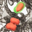
HLOs are grown in a dish and are composed of six types of human stem cells. The multicellular, 3D structures aim to recreate key features of human tissues such as cellular complexity and architecture. They are increasingly used to better understand various pulmonary diseases, from cystic fibrosis to lung cancer, and infectious diseases, including SARS-CoV-2.
Until this study, no one had demonstrated the ability of lung organoids to capture functional pulmonary tissue responses to nanomaterials. To expose the organoid model to carbon-based NMs, lead scientist Rahaf Issa, PhD, developed a method to accurately dose and microinject nanomaterials into the organoid's lumen. It simulated the real-life exposure of the apical pulmonary epithelium, the outermost layer of cells lining respiratory passages within the lungs.
Existing animal research data has shown that a type of long and rigid multi-walled carbon nanotubes (MWCNT) can cause adverse effects in lungs, leading to persistent inflammation and fibrosis. Using the same biological endpoints, the team's human lung organoids showed a similar biological response, thus validating them as tools for predicting NM-driven responses in lung tissue. The human organoids enabled better understanding of interactions of nanomaterials with the model tissue, but at the cellular level.
MWCNT were shown to induce a persistent interaction with alveolar cells, with limited mucin secretion that leads to the growth of fibrous tissue. In contrast, graphene oxide (GO) sheets — flat, thin and flexible forms of carbon nanomaterial — were found to be momentarily trapped out of harm's way in a larger amount of secretory mucin.
Now, Drs. Issa and Vranic, who are based at the university's Center for Nanotechnology in Medicine, are developing and studying a groundbreaking HLO that also contains an integrated immune cell component. This work encourages humane animal research and supports the 3Rs framework of replacement, reduction and refinement, which is established in the U.K. and other countries.
“With further validation, prolonged exposure and the incorporation of an immune component, human lung organoids could greatly reduce the need for animals used in nanotoxicology research,” Dr. Vranic said.
University Chair of Nanomedicine Kostas Kostarelos, PhD, said that although current 2D cell culture models used in NM testing can provide some understanding of cellular effects, they are too simplistic to fully depict the way cells communicate with each other. Therefore, they cannot represent the complexity of the human pulmonary epithelium and may misrepresent the toxic potential of nanomaterials.
“Though animals will still be needed in research for the foreseeable future, 3D organoids nevertheless are an exciting prospect in our research field and in research more generally as a human equivalent and animal alternative,” Dr. Kostarelos said.


